Label the Structures of the Stomach in the Figure. – The Functions of the Stomach
November 9, 2023
Label the Structures of the Stomach in the Figure.
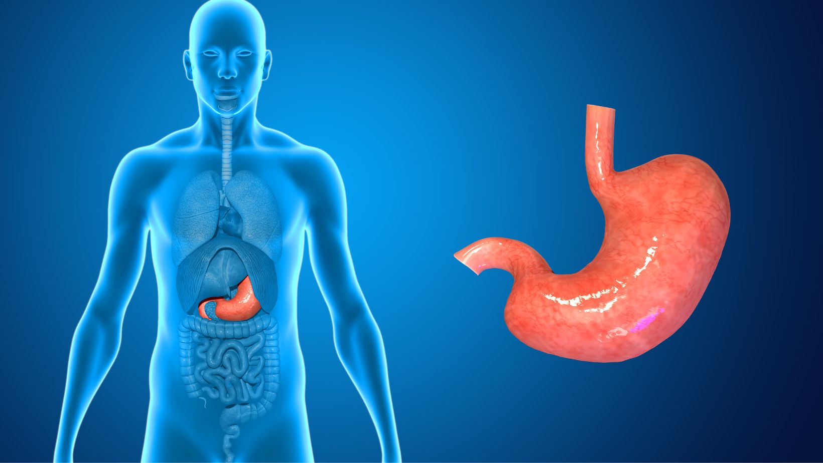
Labeling the structures of the stomach in a figure can be an important exercise in understanding its functions. The stomach, a muscular organ located in the upper abdomen, plays a crucial role in the digestion process. By identifying and naming these structures correctly, we can gain insights into how they contribute to this intricate process.
One key structure of the stomach is the cardiac sphincter, which controls the flow of food from the esophagus into the stomach. This circular muscle relaxes to allow food to enter and then contracts to prevent acid reflux from occurring. Another vital component is the fundus, which acts as a storage area for food before it enters further stages of digestion.
Additionally, we have the body or corpus, where most of the digestive processes take place. It contains gastric glands that secrete enzymes and hydrochloric acid necessary for breaking down proteins and killing bacteria. Lastly, there is the pylorus, responsible for regulating the passage of partially digested food into the small intestine.
Understanding these structures not only helps us visualize how our digestive system operates but also allows us to comprehend their specific roles in maintaining our overall health and well-being. So let’s embark on this journey together as we uncover and explore each component’s function within this fascinating organ!
Structure of the Stomach
Let’s dive into the intricate structure of the stomach and explore its fascinating features. The stomach, a muscular organ located in the upper abdomen, plays a vital role in the process of digestion. It is an integral part of our digestive system, responsible for breaking down food and extracting nutrients that our bodies need to function effectively.
- Shape and Size: The stomach is shaped like a J and can hold approximately 1-2 liters of content. Its size varies depending on factors such as age, body size, and eating habits. The shape allows it to accommodate large amounts of food while maintaining its ability to contract and mix gastric juices with the ingested material efficiently.
- Layers of Tissue: Within the walls of the stomach, several layers of tissue work together harmoniously. These layers include:
- Mucosa: The innermost layer lining the stomach’s lumen consists mainly of epithelial cells that secrete mucus, protecting it from acid damage.
- Submucosa: This layer contains blood vessels and glands that aid in nutrient absorption from digested food.
- Muscularis: Made up of smooth muscles arranged in three layers – longitudinal, circular, and oblique – this layer helps with mixing food through rhythmic contractions.
- Serosa: The outermost layer provides protection against friction by secreting serous fluid.
- Gastric Glands: The gastric glands embedded within the walls produce various substances crucial for digestion. These include hydrochloric acid (HCl) responsible for breaking down proteins and intrinsic factors necessary for vitamin B12 absorption. Additionally, pepsinogen is secreted by these glands which gets converted into pepsin – an enzyme that aids protein digestion.
- Cardiac Sphincter and Pyloric Sphincter: At either end of the stomach lie two important sphincters – the cardiac sphincter and the pyloric sphincter. The cardiac sphincter, located at the entrance of the stomach, prevents gastric acid and partially digested food from flowing back into the esophagus. The pyloric sphincter, situated at the exit of the stomach, regulates the passage of food into the small intestine, ensuring controlled digestion.
Understanding the structure of the stomach helps us appreciate its intricate design and function. As we continue our exploration of this remarkable organ’s functions in subsequent sections, we’ll gain a deeper understanding of how it contributes to our overall digestive process. So stay tuned for more fascinating insights!
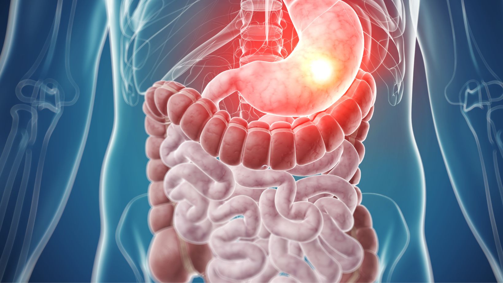
Anatomy of the Stomach
Let’s delve into the fascinating anatomy of the stomach. The stomach, a muscular organ located in the upper abdomen, plays a crucial role in our digestive system. Its intricate structure enables it to carry out its essential functions with remarkable efficiency.
The stomach is divided into four main regions: the cardia, fundus, body, and pylorus. Each region has its distinct characteristics and contributes to different aspects of digestion. The cardia is situated closest to the esophagus and acts as a gateway for food entry into the stomach. The fundus, located above the cardiac region, serves as a reservoir for swallowed food and aids in the initial breakdown through mechanical digestion.
Moving down towards the middle section known as the body of the stomach, we find an abundance of gastric glands that secrete hydrochloric acid and enzymes necessary for chemical digestion. This powerful combination helps break down proteins into smaller molecules that can be absorbed later in our intestines.
Lastly, at the lower part of our stomach lies an important structure called the pylorus. It acts as a valve that regulates passage between our stomach and small intestine by controlling how much partially digested food (chyme) enters at any given time. This controlled release ensures optimal absorption further along in our digestive tract.
To visualize these structures, take a moment to examine Figure X [insert figure here]. You’ll notice how each region is clearly demarcated within this remarkable organ that assists us in processing our daily intake of nutrients.
Amanda Davis
Amanda is the proud owner and head cook of her very own restaurant. She loves nothing more than experimenting with new recipes in the kitchen, and her food is always a big hit with customers. Amanda takes great pride in her work, and she always puts her heart into everything she does. She's a hard-working woman who has made it on her own, and she's an inspiration to all who know her.

How Can a Stay-at-Home Mom Keep the House in a Divorce Setup?
How Can a Stay-at-Home Mom Keep the House in a Divorce As a stay-at-home mom going through a divorce

The Shocking Lee Brady Casey Margenau Fine Homes and Estates, Inc
Lee Brady Casey Margenau Fine Homes and Estates, Inc. is a premier real estate agency known for its

Farmasius Com Login: Access Your Account With Ease
Looking to access your Farmasius account? Look no further than farmasius com login! With this conven